An MRI uses a magnetic field and radio waves to produce detailed images of the body’s internal structures. This technique enables doctors to accurately examine joints and soft tissues for cartilage damage. The cross-sectional images help identify the extent and location of injuries. Here are a few ways an MRI plays a role in identifying cartilage damage:
How MRI Technology Works
MRI uses magnetic fields and radio waves to produce detailed images of internal body structures. The magnetic field realigns water molecules in the body, while radiofrequency signals stimulate these molecules to emit faint signals. MRI sensors detect these signals, which are then processed into detailed images.
The resulting cross-sectional images clearly display cartilage, tendons, ligaments, and other soft tissues. This level of detail helps identify damage that might not be visible with other imaging methods. Unlike X-rays, which primarily reveal bones, MRI provides a clear view of soft tissue structures.
Cartilage Conditions MRI Can Detect
MRI scans provide detailed imaging that helps identify cartilage damage and related joint conditions, allowing doctors to assess the extent of injury and plan appropriate care. The technology can reveal areas of cartilage wear, tears, or degeneration, providing a comprehensive view of joint health.
Cartilage conditions MRI can detect include:

- Cartilage tears
- Cartilage degeneration
- Joint surface damage
- Cartilage defects or lesions
- Early signs of cartilage wear
The MRI Scanning Process
Preparing for a scan involves removing any metal items that could interfere with the imaging process. This includes jewelry, hairpins, underwire bras, and eyeglasses. Removing these items helps produce precise and accurate images.
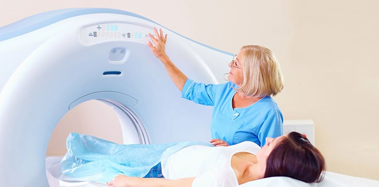
The machine is a long, narrow tube with a sliding table on which patients lie during the scan. Medical staff monitor the procedure from an adjacent room while the machine generates a magnetic field and directs radio waves at the body. The scan typically takes 45 minutes to an hour, is painless, and may include medication for patients who experience claustrophobia, though the machine produces loud knocking sounds during operation.
Benefits of In-Office MRI Services
In-office MRI services simplify the diagnostic process by allowing patients to receive imaging and consultation at the same location, reducing wait times and scheduling challenges. Medical staff often coordinate with insurance providers to obtain authorization before the scan, helping patients understand coverage and potential costs. This setup enables prompt discussion of results and treatment planning, making the overall process more efficient and less stressful.

What to Expect After Your MRI
After a scan, doctors review the images to identify cartilage damage and related conditions. Patients typically discuss the results shortly after the imaging is complete. Unless sedatives were used, they can return to normal activities immediately without restrictions.
The detailed images provide doctors with comprehensive information for developing treatment strategies. Depending on the severity and location of the cartilage damage, options may include physical therapy, lifestyle adjustments, or surgical intervention. This information helps guide the next steps in care and monitoring.
Making Informed Decisions About MRI
MRI technology offers a safe and non-invasive method for evaluating cartilage damage and examining soft tissue structures in detail. The images help healthcare providers distinguish between different types of injuries and guide treatment decisions. Individuals experiencing joint pain, limited mobility, or suspected cartilage issues may discuss MRI options with their provider to better understand their condition.