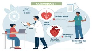
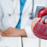
Introduction
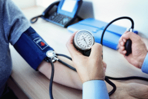
Cardiologists rely on a variety of diagnostic tests to assess the structure and function of the heart. These tests help detect conditions like coronary artery disease, arrhythmias, heart failure, and valve disorders. Choosing the right test depends on the patient’s symptoms, medical history, and risk factors. Understanding these tests can help patients feel more informed and prepared during their cardiology visits.
Electrocardiogram (ECG or EKG)
An electrocardiogram is one of the most basic and commonly used tests. It records the electrical signals of the heart to identify:
- Abnormal heart rhythms (arrhythmias)
- Signs of a past or current heart attack
- Changes in heart muscle thickness
This test is quick, painless, and provides valuable initial information about heart function.
Echocardiogram
An echocardiogram uses ultrasound waves to create real-time images of the heart. It helps cardiologists evaluate:
- Heart size and shape
- Pumping strength (ejection fraction)
- Valve function
- Fluid buildup around the heart
It’s often performed when there are symptoms like shortness of breath or heart murmurs.

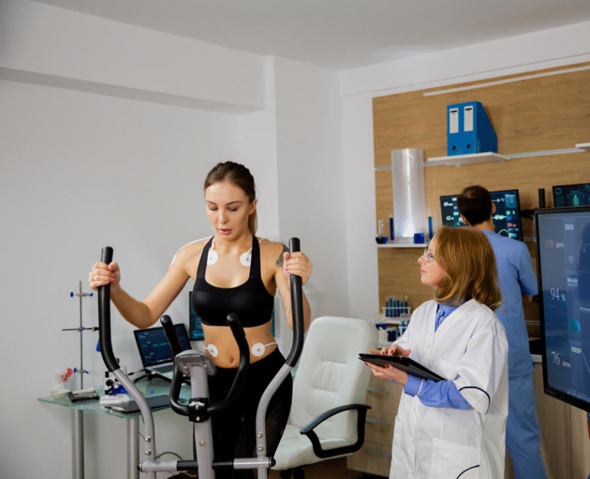
Stress Test
A stress test shows how the heart responds to exertion. The most common type involves walking on a treadmill while heart activity is monitored. It helps detect:
- Reduced blood flow to the heart
- Exercise-induced arrhythmias
- Cardiovascular fitness
In patients who cannot exercise, medications are used to simulate stress on the heart.
Holter Monitor
A Holter monitor is a portable device worn for 24 to 48 hours that records heart rhythms continuously. It is useful for detecting:

- Intermittent arrhythmias
- Heart rate variability
- Palpitations not captured during clinic visits
Patients wear the monitor while going about normal daily activities.
Event Monitor
Similar to a Holter monitor, an event monitor is worn for longer periods—sometimes up to 30 days. It is activated by the patient when symptoms like dizziness or chest pain occur. It provides detailed rhythm analysis over time, especially when symptoms are infrequent.
Cardiac Catheterization and Angiography
Cardiac catheterization is an invasive test that involves threading a thin tube through blood vessels to the heart. Dye is injected to make arteries visible on X-rays. This test helps diagnose:
- Blocked or narrowed coronary arteries
- Valve disease
- Heart function and chamber pressures
It often guides treatment decisions like stent placement or surgery.
Cardiac MRI
Magnetic resonance imaging (MRI) provides highly detailed images of the heart’s structure. It helps assess:
- Tissue damage after a heart attack
- Congenital heart defects
- Inflammation or scarring
It is non-invasive and useful when echocardiogram results are unclear.
Cardiac CT Scan
A cardiac CT scan uses X-rays to create 3D images of the heart and blood vessels. It is especially helpful for:
- Detecting coronary artery disease
- Visualizing calcium buildup (calcium score test)
- Evaluating heart anatomy before surgery
It provides rapid and accurate data, especially for low-risk patients.
Blood Tests
Various blood tests are used to support a cardiac diagnosis. These include:
- Troponin (for heart attack detection)
- BNP (for heart failure)
- Lipid profile (cholesterol levels)
- C-reactive protein (inflammation marker)
Blood tests offer insight into heart muscle damage and risk factors for cardiovascular disease.
Tilt Table Test
This test is used when patients experience unexplained fainting. The patient is strapped to a table that tilts to change position, while heart rate and blood pressure are monitored. It helps diagnose:
- Low blood pressure
- Nervous system disorders affecting circulation
- Certain arrhythmias
It provides useful data for understanding syncope (fainting episodes).
Nuclear Stress Test
This advanced stress test involves injecting a small amount of radioactive material to assess blood flow to the heart muscle. A special camera captures images during rest and stress phases. It is used to:
- Detect blockages
- Assess heart damage after a heart attack
- Guide treatment decisions
It provides greater detail than a standard stress test.
Electrophysiology Study (EPS)
An EPS is used to study the electrical system of the heart in patients with serious arrhythmias. Thin wires (catheters) are inserted into the heart to map its electrical activity. It helps locate the source of abnormal rhythms and guide treatments like ablation.
Conclusion
Cardiology uses a broad range of tests to diagnose and monitor heart conditions—from simple ECGs to advanced imaging and invasive studies. Each test provides specific information about heart rhythm, structure, blood flow, or function. Early and accurate testing helps cardiologists design effective treatment plans and reduce the risk of serious complications. Understanding these tests allows patients to be better informed and more active in their own care.
FAQs
Which heart test is most commonly used?
The electrocardiogram (ECG) is the most frequently used initial test to detect heart rhythm and damage.
Are heart tests painful?
Most tests are non-invasive and painless. Some invasive tests like catheterization involve mild discomfort but are done under local anesthesia.
How long does a stress test take?
A standard exercise stress test usually takes about 30–45 minutes, including preparation and recovery.
What test checks for blocked arteries?
Cardiac catheterization and nuclear stress tests are used to check for blocked or narrowed arteries.
When should I ask for heart testing?
If you have chest pain, shortness of breath, dizziness, or a family history of heart disease, consult your doctor for appropriate testing.